Scientists Map 539 Distinct Chemical Zones in Mouse Brain
At a Glance
- Researchers have mapped the distribution of lipids across the mouse brain.
- The atlas identifies 539 chemically distinct regions, revealing previously overlooked patterns.
- Findings could help scientists investigate brain aging, pregnancy, and neurological disorders.
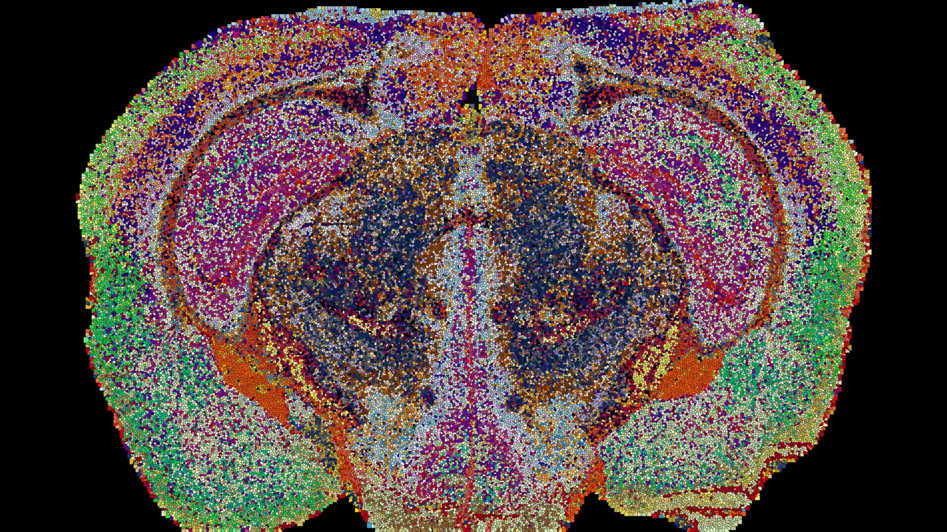
Researchers at Switzerland’s Ecole Polytechnique Federale de Lausanne (EPFL) have created a detailed three-dimensional map of lipids across the mouse brain, identifying 539 chemically distinct regions that could offer new insights into brain organization, pregnancy, and diseases such as Alzheimer’s.
Key Statements and Focus Area
- Brain Organization: “In effect, lipids work like postal codes, giving distant but related parts of the brain the same chemical address.” — Giovanni D’Angelo, EPFL
- Research Significance: The atlas reveals chemical differences that may not be apparent through conventional anatomical or genetic mapping.
- Focus Area: A new lipid mapping method could help researchers study changes in brain chemistry during development, aging, and disease.
Mapping 539 Chemical Regions
The research team, led by Giovanni D’Angelo, Luca Fusar Bassini, and Gioele La Manno, used mass spectrometry imaging to examine the distribution of lipids in mouse brain tissue. The findings were published in the scientific journal Nature.
The researchers analyzed 172 lipid types across 109 brain slices collected from 11 mice, generating approximately seven million measurements. They combined the data with machine learning tools to build a three-dimensional reconstruction of the brain’s lipid distribution.
The resulting map identified 539 chemically distinct regions, which the team named “lipizones.” Each region has a characteristic lipid composition, allowing researchers to distinguish areas that may appear similar using conventional mapping methods.
Lipids are fatty molecules that form cell membranes, contribute to the myelin insulation surrounding nerve fibers, and help regulate communication between brain cells. Despite their importance, their precise distribution across the brain has been difficult to map.
Hidden Differences in Brain Tissue
The atlas revealed that some lipid patterns correspond to established brain structures, while others identify chemical boundaries that previous approaches had overlooked.
White matter, which contains nerve fibers connecting different brain regions, showed particularly diverse lipid compositions. The findings suggest that cells responsible for producing myelin may have greater chemical variation than genetic maps alone indicate.
Researchers also identified previously unrecognized chemical boundaries in the choroid plexus, which produces cerebrospinal fluid, and in tissues lining the brain’s internal cavities.
The team found that lipid signatures could help identify the original location of a tissue sample, suggesting that lipid composition provides another way to understand the brain’s organization.
Pregnancy Changes Brain Lipid Composition
The researchers also used the atlas to examine changes in the brains of pregnant mice. They found widespread shifts in lipid composition across multiple regions, with changes more pronounced overall than typical differences between male and female mice.
The cortex showed particularly extensive changes, while levels of galactosylceramide, a lipid important for myelin, increased sharply across much of the brain’s white matter.
The researchers said the findings suggest that pregnancy is accompanied by substantial changes in brain chemistry. Further research will be needed to establish how these changes affect neural function and adaptation during pregnancy.
Potential Applications in Brain Disease Research
The Lipid Brain Atlas is freely available online, giving researchers access to a reference map of lipid distribution in healthy mouse brain tissue.
By combining lipid maps with existing genetic and cellular data, scientists may be able to investigate how brain chemistry changes during development and aging and how disruptions could be associated with conditions such as Alzheimer’s disease and depression.
The researchers cautioned that the mouse atlas cannot simply be applied to the human brain. However, the mapping method could be adapted to investigate lipid organization in human brain tissue.
FYI
Lipids are essential components of the nervous system. They help maintain cell membranes, support the myelin sheaths that insulate nerve fibers and contribute to signaling between cells. Differences in lipid composition can therefore provide clues about how brain regions function and how their biology changes.
The atlas adds a chemical layer to existing maps of brain anatomy, genes, and cell types. Its future value will depend on how researchers use it to investigate specific biological processes and compare healthy tissue with tissue affected by disease.
Dive Deeper: Handpicked Stories for You
Follow Channel8 for continuous updates:
2 hours ago